Floor Of Third Ventricle Is Formed By
Third Ventricle Anatomy Kenhub
Third Ventricle Medfriendly Com
Third Ventricle Assignment Point

Third Ventricle Location Boundaries Recesses And Choroid Plexus Anatomy Qa
Csf And Ventricular System Ppt Video Online Download

The Third Ventricle Ventricles Of The Brain
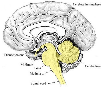
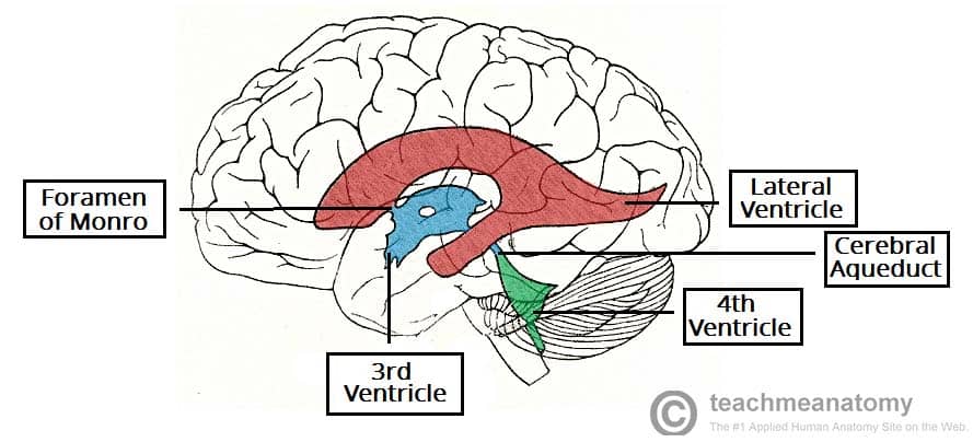
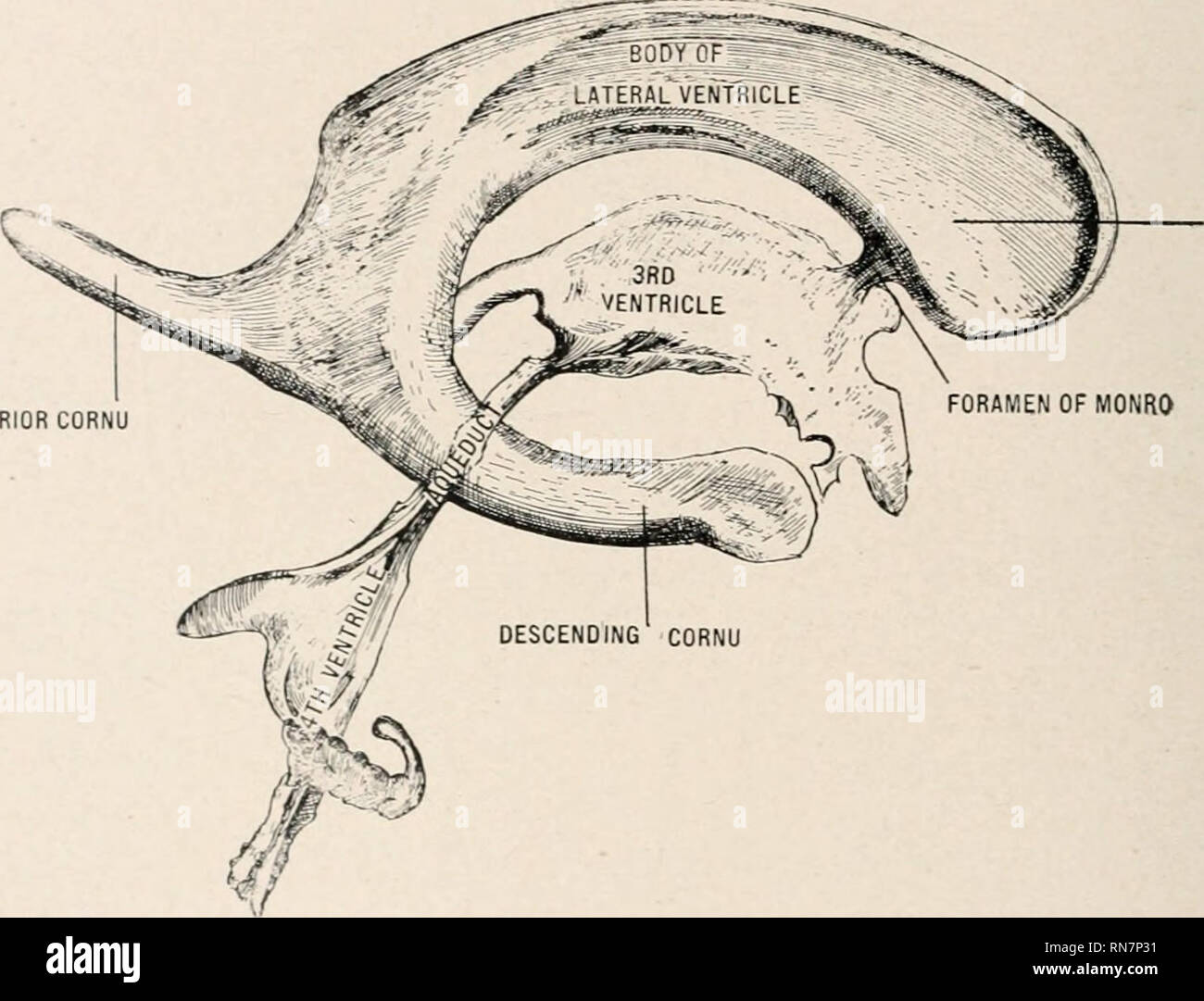
The third ventricle is located directly above the midbrain.
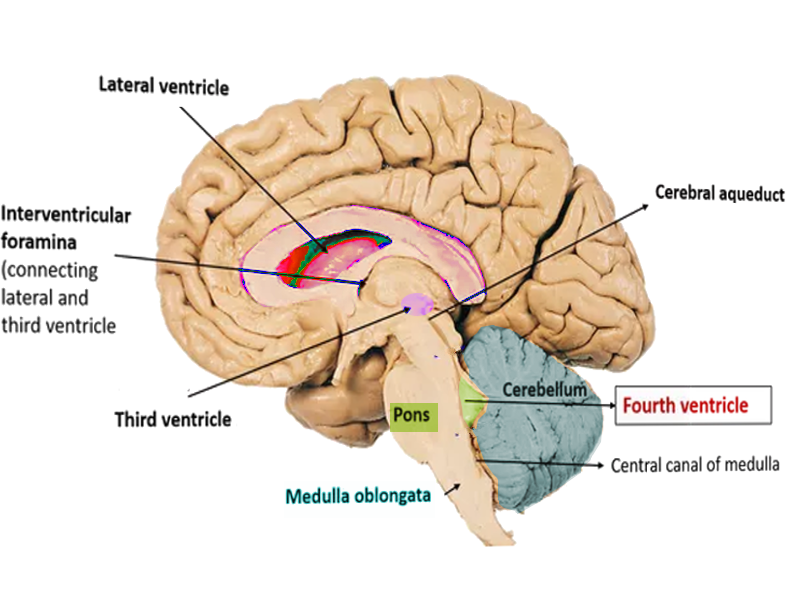
Floor of third ventricle is formed by. The third ventricle in blue is a narrow vertical cleft between the 2 lateral ventricles it possesses a roof a floor and four walls. You can see how the top of the midbrain forms part of the floor of the third ventricle. The third ventricle is a narrow midline cavity that communicates through the foramen of monro with the lateral ventricles and through the aqueduct with the fourth ventricle. The third ventricle has.
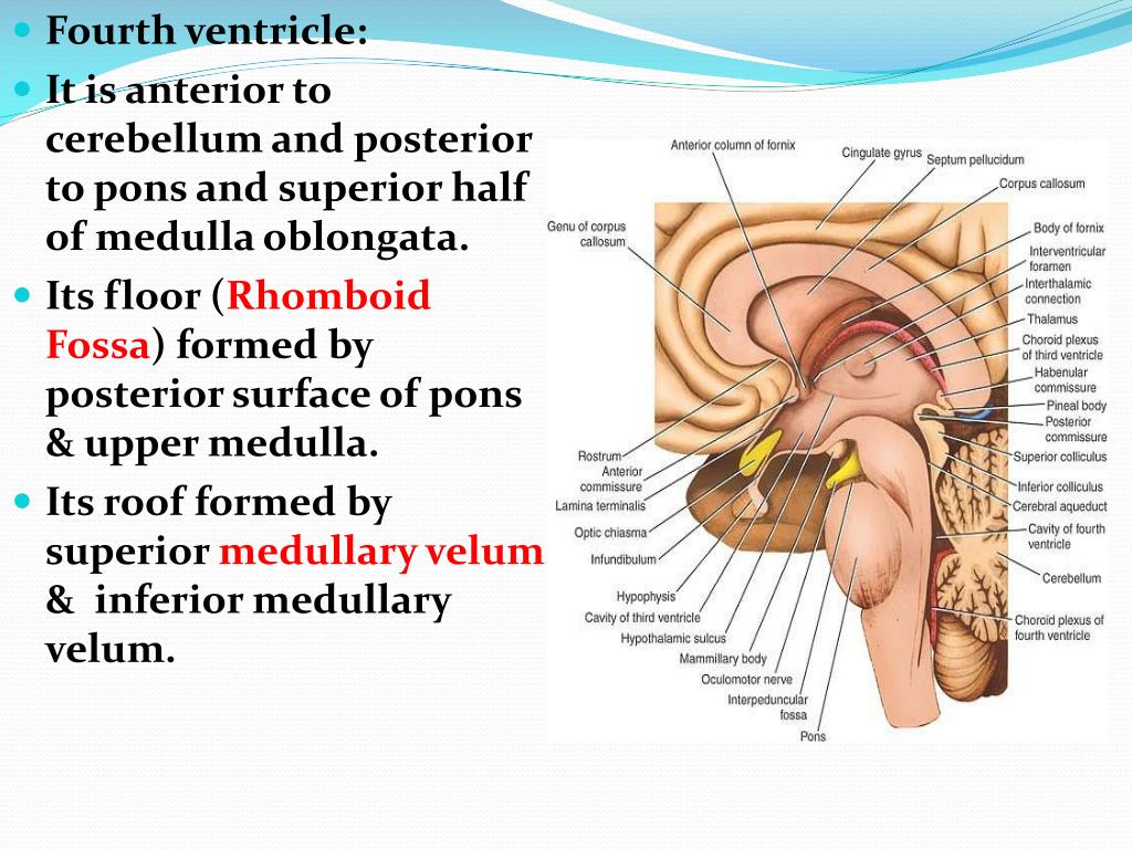
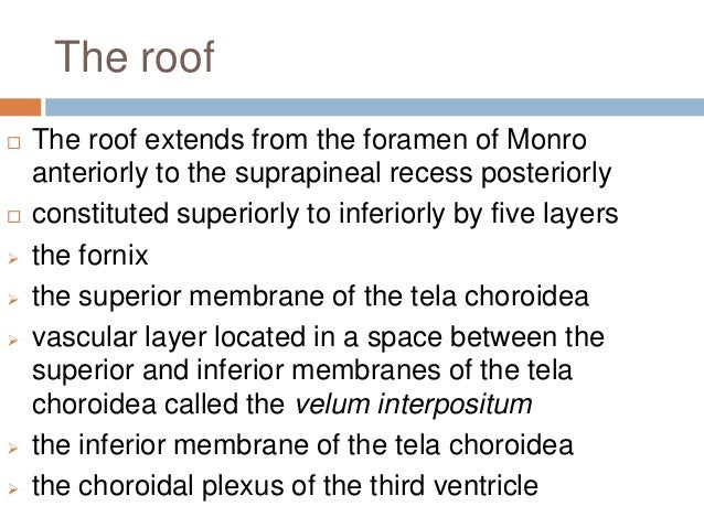
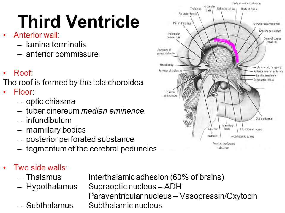
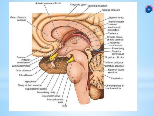
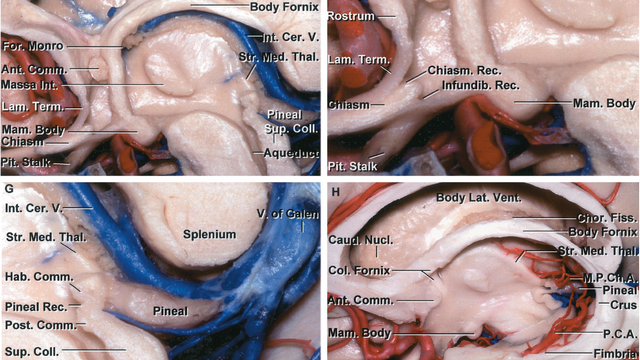
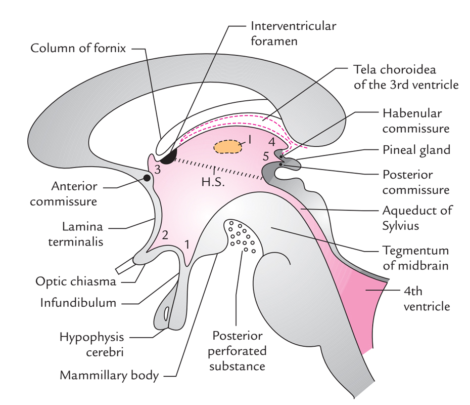
The roof is formed by the tela choroidea. The lateral walls of the third ventricle are formed by the walls of the left and right thalamus. The floor of the third ventricle is formed by a number of structures including the hypothalamus subthalamus mammilary bodies infundibulum pituitary stalk and the tectum of the midbrain. The third ventricle is one of the four connected ventricles of the ventricular system within the mammalian brain it is a slit like cavity formed in the diencephalon between the two thalami in the midline between the right and left lateral ventricles and is filled with cerebrospinal fluid csf.
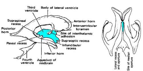
The inferior part is formed by the inferior cerebellar peduncle and by the gracile and cuneate tubercles of the brainstem. The third ventricle has two lateral walls a roof a floor an anterior and a posterior wall. It has two major extensions known as the lateral recesses one on either side of the midline. Roof formed by body of fornix and the ependyma lining the under surface of the tela choroidea.
The lateral walls of the fourth ventricle are formed by the cerebellar peduncles. Running through the third ventricle is the interthalamic adhesion which contains thalamic. The floor of the third ventricle is ormed by optic chiasma the infundibulum and the tubular cinereum. Like other ventricles the third ventricle has a cavity an anterior wall a posterior wall a floor a roof and two lateral walls.
The superior part of these walls is formed by the superior cerebellar peduncle. Formed by four layers. It is a cavity within diencephalon it is a midline slit like cavity situated between the two thalami and the part of hypothalamus. Lamina terminalis roof.
The narrow roof of the third ventricle is formed by. Anterior wall is formed from above downwards by. Floor of third ventricle formed primarily by hypothalamic structures optic chiasma infundibular recess which extends into pituitary stalk tuber cinereum mammillary bodies. Anterior posterior and two lateral.
The Ventricles Of The Brain Lateral Third Fourth Teachmeanatomy
Meninges Csf Ventricular System Objectives Describe The Arrangement Of The Meninges And Their Relationship To Brain And Spinal Cord Explain The Occurrence Ppt Download
Third Ventricular Surgical Approaches

Ventricles Functions Flashcards Quizlet
Midsagittal Views Of The Third Ventricle Neuroanatomy The Neurosurgical Atlas By Aaron Cohen Gadol M D
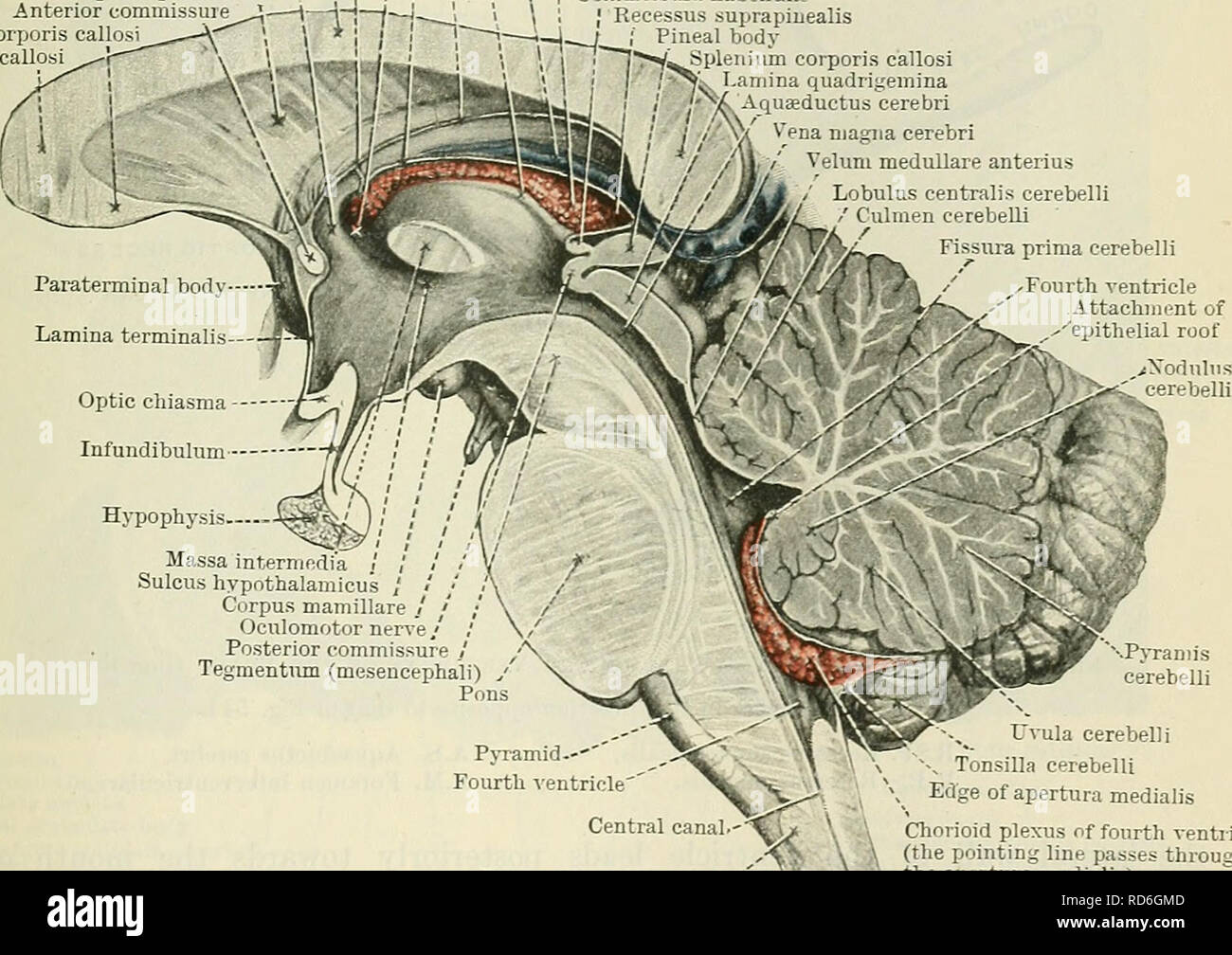
Cunningham S Text Book Of Anatomy Anatomy Paets Derived From The Diencephalon 617 Matter Which Surrounds The Aqueduct Is Directly Continuous With The Gray Matter Of The Substantia Perforata Posterior And Tuber Cinereum
Neuroanatomy Fourth Ventricle Article Statpearls
Third Ventricle Location Anatomy Function Features
Third Ventricle Surgical Anatomy And Approaches

Diencephalon And Third Ventricle Neupsy Key

Brain 101 The Ventricles And Csf Flow Hydrocephalus Association

Schematic Drawing Of The Floor Of The Third Ventricle Ideally The Download Scientific Diagram
Anatomy In A Nutshell A Treatise On Human Anatomy In Its Relation To Osteopathy Human Anatomy Osteopathic Medicine Osteopathic Medicine Anatomy 352 Anatomy In A Nutshell Choroid Plexus Of The